
A seborrheic keratosis (seb-o-REE-ik ker-uh-TOE-sis) is a common benign skin growth, similar to a mole. Most people will have at least one in their lifetime. They tend to appear in mid-adulthood and their frequency increases with age. They are harmless and don’t require treatment, but you can have them removed if they bother you.
Contents
- 1 What is seborrheic keratosis?
- 2 What does seborrheic keratosis look like?
- 3 Who gets seborrheic keratosis?
- 4 Symptoms and Causes
- 5 Do seborrheic keratosis cause symptoms?
- 6 If these symptoms annoy you, you may want to have the growth removed.
- 7 How do you tell seborrheic keratosis from actinic keratosis?
- 8 Case Presentation
- 9 Discussion
What is seborrheic keratosis?
Skin growths like seborrheic keratoses are sometimes also called epidermal tumors. That doesn’t mean they’re cancer, though. Technically, moles and warts are also epidermal tumors. That just means they are clusters of extra cells on the epiderma, the outer layer of the skin. They aren’t considered a risk factor for skin cancer.Paraneoplastic diseases may be defined as hormonal, neurological, or hematological disturbances and as clinical and biochemical imbalances related to the presence of malignancies, with no direct association with primary tumor invasion or metastasis . If promptly recognized, skin manifestations can lead to an early diagnosis of neoplasia, improving its prognosis .
Among the cutaneous findings is the rare and controversial sign of Leser-Trélat, characterized as the sudden appearance and/or increase in number and size of seborrheic keratoses, associated with an internal neoplasm. The aim of this case report is to describe a cancer diagnosis made after following the clue given by the patient’s skin condition—a case of Leser-Trélat syndrome.
What does seborrheic keratosis look like?
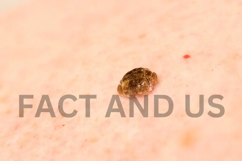
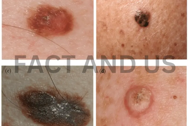
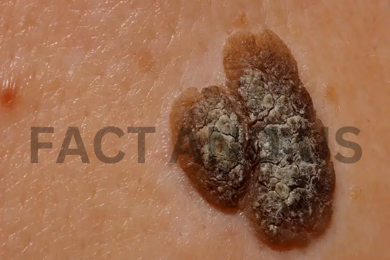
Seborrheic keratoses are roundish or oval-shaped patches on the skin with a “stuck on” appearance. They are sometimes described as waxy or scaly. They are raised above the skin and even when they are flat you can feel them with your finger. They are usually brown, but can also be black or tan, and less often, pink, yellow or white. They often appear in numbers.
Seborrheic keratoses are characterized by keratin on the surface — the same fibrous protein that fingernails, hooves, and horns are made of. This causes the textural details that often distinguish the growths. Sometimes it looks like small bubbles or cysts within the growth. Sometimes it looks scabby or wart-like. Sometimes it looks like the ridges and fissures in a brain.
Who gets seborrheic keratosis?
Anyone can get one, but most commonly:
- People who are 50 and older. They usually begin to appear in middle age, and rarely in younger people. About 30% of people have at least one by the age of 40, and about 75% by the age of 70.
- People with a family history of it. About half of all cases of multiple seborrheic keratoses occur in mfamilies, suggesting that the tendency to develop many of them may be inherited.
- Lighter-skinned people. Classic seborrheic keratosis as described here appears less frequently in darker-skinned people. However, a variant of seborrheic keratosis called dermatosis papulosa nigra is very common in darker-skinned people, including those of African, Asian and Hispanic descent.
Symptoms and Causes
What triggers seborrheic keratosis?
We don’t know exactly why these growths occur, but we can look at the circumstances that often go along with it. The first is age: seborrheic keratoses are especially common in adults over 50, and they tend to multiply as people get older. Some studies suggest that sun exposure may increase their occurrence. They also appear more frequently in families, which suggests that genetics may play a role. They are not viral or bacterial. They don’t spread and they aren’t contagious.
Seborrheic keratoses usually grow slowly and may develop their texture gradually over time. If many seborrheic keratoses erupt suddenly together, it might raise some concern. This unusual occurrence has sometimes been considered a sign of internal cancer. Doctors call it “the sign of Leser Trélat”. The correlation is not yet proven or explained and may only be a coincidence. But your healthcare provider might want to screen you for any other signs of internal cancer.
Do seborrheic keratosis cause symptoms?

They usually don’t, but people sometimes report:
- Itching.
- Irritation from friction.
- Bleeding.
If these symptoms annoy you, you may want to have the growth removed.
Diagnosis and Tests
How do you tell seborrheic keratosis from actinic keratosis?
Seborrheic keratosis and actinic keratosis can resemble each other. They both begin to appear after the age of 40, and they both can appear crusty and scaly. It’s important to know the difference because actinic keratosis is more serious than seborrheic keratosis. Unlike seborrheic keratosis, actinic keratosis is caused by sun exposure, and it carries a small risk of turning into skin cancer (5% -10%).
Actinic keratoses vary in color, but they tend to be less pigmented than seborrheic keratoses. They can be flat or slightly raised, but tend to be flatter, and you might feel them before you see them. They feel scaly and rough, but may become more bumpy and wart-like over time, like seborrheic keratoses. They may also itch and bleed. They tend to appear in clusters in frequently sun-exposed areas.
Case Presentation
A 67-year-old man was admitted to the intensive care unit (ICU) due to a second exacerbation of COPD in two weeks. He came from a first aid unit, had already been through orotracheal intubation, and was receiving mechanical ventilation and antibiotics.
The patient was a heavy smoker (120 pack-years of tobacco exposure) and started to claim dyspnea to great efforts over the previous few months; he had also lost 8 kg in less than a month. There was no other known comorbid condition in his clinical history. After some days of stabilization in the ICU, the patient was referred to the general internal medicine unit, where another exacerbation took place, along with fever and productive cough, likewise controlled with bronchodilators and antibiotics.
On physical examination, multiple seborrheic keratoses on the back of the hands , elbows, and trunk were observed. They were asymptomatic and have appeared in the previous four months. These lesions alerted to the possibility of a paraneoplastic manifestation, hypothesis also considered by the dermatologists who followed the case. The search for an occult neoplasia was then initiated. The investigation comprehended thorax CT-scan, upper gastrointestinal endoscopy, colonoscopy, and blood analysis, which pointed no malignancy.
Colonoscopy showed four sessile polyps, one in the ascending colon (measuring 10 mm), histopathologic ally defined as a tubulovillous adenoma with high-grade dysplasia, and three in the transverse colon (the biggest measuring 9 mm), corresponding to tubular adenomas with high-grade dysplasia. Ultimately, abdomen CT-scan revealed one irregular-shaped vegetation on the bladder floor, measuring 22 × 18 × 16 mm, with no apparent adipose tissue invasion . The patient denied previous hematuria or any other urinary symptom.
Discussion
Indirect involvement of the skin by visceral tumors can cause a variety of inflammatory, proliferative, metabolic, and neoplastic changes without the actual presence of tumor cells . In these paraneoplastic disorders, the skin condition generally shows up distant from the primary tumor site. Though mechanisms are unknown in most cases, it is believed that mediators such as growth factors, cytokines, or hormones are involved in the pathogenesis of the cutaneous findings .
Six criteria have been described to consider a cutaneous alteration as paraneoplastic, known as Curth postulates: both conditions (neoplasia and paraneoplasia) are of concurrent onset, both follow a parallel course, the skin lesion is not part of a genetic syndrome, a specific tumor occurs with a certain dermatosis, the dermatosis is not common in general population, and there is a high percentage of the association between both conditions . Not all criteria must be met to demonstrate the link between a skin disease and an underlying malignancy .
The sign of Leser-Trélat is defined as the sudden appearance and/or rapid increase in number and size of seborrheic keratoses, secondary to a neoplasia . These are papular, verrucous, usually well-defined lesions of varying colors (brown, black, or tan) which primarily affect thorax and dorsum, followed by extremities, face, abdomen, neck, and axilla . Though the description of the sign has been credited to Edmund Leser and Ulysse Trélat, they were both observing the presence of cherry angiomas in oncological patients, association that probably does not exist; Hollander was indeed the first to document the connection between cancer and eruptive seborrheic keratoses.
In 1988, Holdiness gathered the available data (60 cases) to analyze clinical features of the sign. The average age at the time of reported onset was 60.7 years. The average time of evolution of the cutaneous lesions was 14.9 weeks, highlighting its eruptive presentation as a requisite that get most of the patients to be aware of. Acanthosis nigricans and pruritus were simultaneously present in 16.7%. Of these patients, 55% had adenocarcinomas, of which 36.4% were adenocarcinomas of the stomach; lymphoproliferative neoplasm were the second most recorded neoplasms, accounting for 18.3%. Metastatic disease was reported in 56.7% of the patients, with an average survival time of 11.5 months after cancer diagnosis .
Stay connected with Fact and US for more such news.